show your support
Cherry Eye
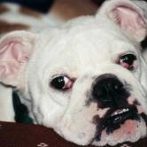
One of the most common eye conditions in Bulldogs.
The gland of the third eyelid is located within the base of the third eyelid. The third eyelid lies between the lower eyelid and the eyeball at the inside corner of each eye. The third eyelid protects the eye and helps contain and distribute tears over the surface of the eye. The third eyelid gland is a tear gland. A prolapse of the gland of the third eyelid results in a pink to red mass in the inner corner of the eye. This is commonly referred to as Cherry Eye. The cause for the prolapse is believed to be partially due to a weakness of the connective tissue attachment of the gland. The tear duct becomes blocked and obstructs tear flow. After a couple of days it begins to swell because now an infection has begun.
Restoring the gland to its normal position will preserve tear production, enhance appearance, and prevent corneal and conjunctival disease caused by the prolonged exposure. Surgically replacing the gland to its normal position is especially important in breeds that are also prone to develop tear insufficiency disease.
Treatment Options:
The most successful surgical technique is called the Conjunctival Mucous Pocket Procedure which creates a pocket to place the prolapsed gland inside and then the pocket is closed with sutures. Another method is to merely suture the gland with sutures (or tack it down). Another procedure, which we do not recommend, is the removal of the gland. This procedure often leaves your pet with very inefficient tear flow which can lead to the serious problem of dry eye, and can result in permanent corneal damage, drops will be required to be placed in the dogs eyes for the rest of his life when removal is done.
Some swelling and redness of the eyelid and lower eyelid is normal after surgery. Keep the eye clean of discharges. Please restrict your pet's activity for the first 2 weeks after surgery to aid in retention of the gland.
An antibiotic and/or anti-inflammatory eye medication is prescribed to try and prevent infection and to minimize inflammation.
More articles we recommend:

Hot Content


Sign up for our free newsletter
Recently made posts
-
Hi everyone! I'm just wondering if anyone is still here!by...1 year 30 weeks ago
-
Hi to everyone! Griffin and I are doing well. A new problem has come up where he won...by Carol...2 years 51 weeks ago
-
Hope you guys are well!by Kathy...4 years 29 weeks ago
-
Happy holidays if anyone is on here!by Kathy...4 years 45 weeks ago
-
This is Carol and Griffin. We've been absent for awhile. It was so fun to see so...by Carol...5 years 2 days ago